
Practical pulse oximetry for the Veterinary Nurse
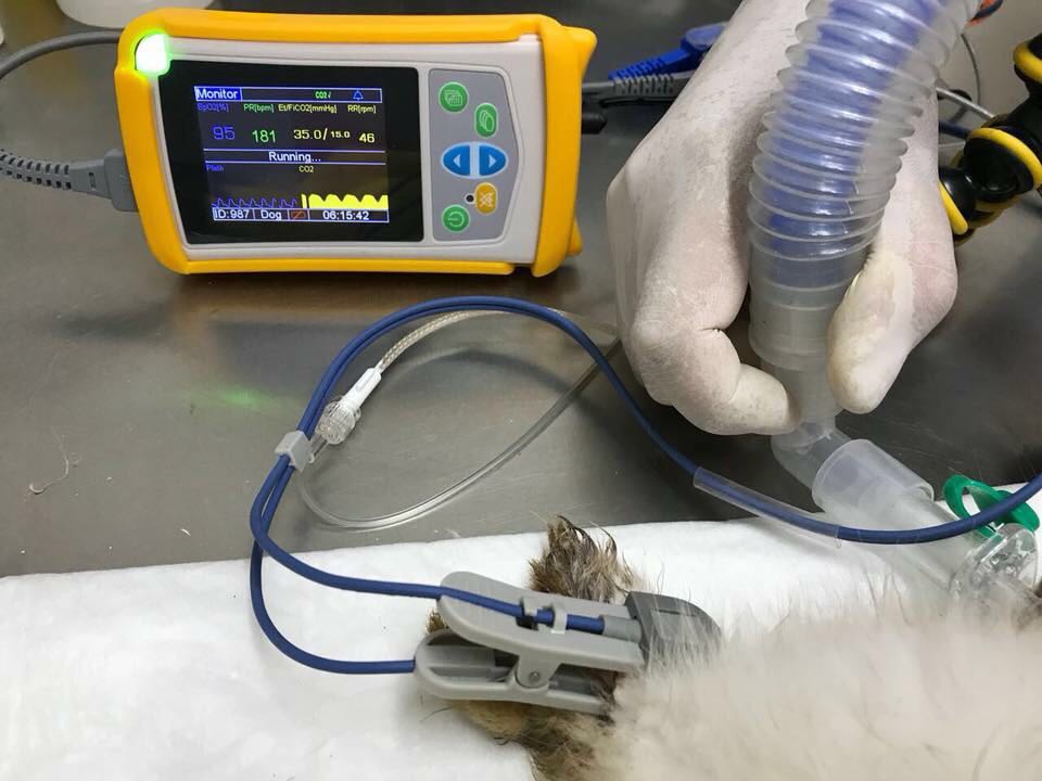
Pulse Oximetry and OxygenationIntroductionPulse oximeters are a widely available piece of anaesthesia patient monitoring equipment used in veterinary practices. They are inexpensive, easy to use, and provide a non-invasive method to gain real-time information on oxygen transport, accompanied by an audible pulse rate. The use of pulse oximetry has been shown to reduce the risk of anaesthesia related deaths (Brodbelt et al., 2008). Oxygen TransportOxygen is transported from the lungs into the blood where it is released into the tissue for cellular metabolism. Oxygen diffuses into the plasma and then binds to the protein haemoglobin found in red blood cells, however, a very small amount of oxygen remains dissolved in plasma. Each red blood cell carries millions of haemoglobin molecules; when the four subunits of the haemoglobin molecule are each carrying an oxygen molecule, the haemoglobin become ‘saturated’ and is referred to as “oxyhaemoglobin”, conversely when haemoglobin is not carrying oxygen it is referred to as “deoxyhaemoglobin”. The pulse oximeter uses a light absorption technique to determine how much of the haemoglobin in arterial blood is saturated with oxygen (SpO2). The Pulse Oximeter ProbeThe most common probe used is the transmission probe; this clip design holds a thin piece of tissue between it and emits an infrared light and a red light from one side of the clip to a receiver on the other side. Placement of the probe should be on pigment free tissue where contact can be made: mucous membranes like the tongue and lip are commonly used, however the prepuce, vulva, ear or webbing between the toes can be used too. If the patient has dark or thick fur, readings may be very difficult to obtain or they may be inaccurate. The probe selectively reads the (pulsatile) arterial blood flow of the tissue; oxyhaemoglobin absorbs more infrared light and deoxyhaemoglobin absorbs more red light at different wavelengths. The pulse oximeter then gives a reading based on the difference between these two absorbances. For example, if an SpO2 reading is 97%, it means that 97% of the red blood cells that were read by the probe had haemoglobin molecules that were fully saturated with oxygen, and 3% were not.
What is Pulse Oximetry telling us?Pulse Oximetry provides information on oxygen transport; its relationship to the Oxyhaemoglobin Dissociation Curve and the Partial Pressure of Arterial Oxygen (PaO2) is used to presume there are normal levels of oxygen in the blood for cellular metabolism in the tissues. In a healthy patient breathing room air, a normal PaO2 is 80-100mmHg which correlates to an SpO2 of 95%-99%. The PaO2 measures the amount of oxygen that is dissolved in plasma after the red blood cells are fully saturated. PaO2 is what drives the diffusion of oxygen into the tissues, so think of SpO2 as the oxygen reservoir until it is released into the plasma. Hypoxemia is defined as a PaO2 is less than 60mmHg which correspond to an SpO2 <90% (Schauvliege, 2016), and may occur during anaesthesia for the following reasons:
Reliability and LimitationsWhen validating an SpO2 reading, the plethysmograph should have a strong waveform which mimics an arterial blood pressure trace and it should also match the heart rate. The upward wave represents systole (the heart contracting) and the downwards wave represents diastole (when the heart is filling before the next beat). A dicrotic notch can be seen on the downwards wave when the aortic valve closes and the distended aorta contracts, creating a brief change in pressure into the arterial circulation. Both the waveform and dicrotic notch can be interpreted alongside other parameters to understand the patient's cardiovascular status:  Pulse oximetry provides a reading based on the red blood cells present as they pass the probe sensors. A limitation in SpO2 readings occurs in anaemic animals where there are fewer red blood cells but they are fully saturated giving a normal SpO2 , but there is still a reduced oxygen carrying capacity overall because there is less haemoglobin. Haemoglobin is estimated as ⅓ of the packed red blood cell volume (haematocrit) and is normally 12.1-20.3g/dL in dogs, and 9.3-15.9g/dL in cats (Klaassen, 1999). Even if both the SpO2 and PaO2 are normal, it does not always indicate that the Oxygen Content of Arterial Blood (CaO2) is normal. To fully assess the oxygen carrying ability of the patient, the haemoglobin levels must be measured. A normal CaO2 in dogs has been reported between 14.2-17.8mL/dL (Haskins et al., 2005) and can be calculated as follows: CaO2 (mL/dL) = (1.36 × [Hb] × % saturation) + (PaO2 × 0.003) To estimate the PaO2 , the FiO2 is multiplied by five; e.g. room air is 21% oxygen and when multiplied by five, the expected PaO2 is 80-100mmHg. In a patient under general anaesthesia breathing 100% oxygen, the PaO2 should be 400-500mmHg. A limitation of pulse oximetry when the patient is breathing an oxygen rich concentration (high levels of PaO2 are achieved), is that the SpO2 will read 99-100%. Therefore, the PaO2 must fall to a dangerously low level for there to be noticeable changes in the SpO2 reading in this instance. A small change in the SpO2 percentage actually reflects a huge change in PaO2. Desaturation (SpO2 <90%) can occur within 60 seconds in dogs breathing room air, compared to 300 seconds in those that were preoxygenated for 3 minutes prior to anaesthesia induction (McNally, Robertson and Pablo, 2009) despite a PaO2 that would have been rapidly decreasing. Two other forms of haemoglobin exist but they do not carry oxygen;
As mentioned previously, pulse oximetry provides information on oxygen transport, not oxygen delivery to the tissue. Oxygen delivery is determined by cardiac output and haematocrit (available haemoglobin). Additionally, it does not provide information on ventilation, and a hypoventilating patient under anaesthesia breathing 100% oxygen can have a normal SpO2 but a dangerously high level of CO2. Troubleshooting abnormal readingsAlthough there are few limitations with pulse oximetry use, inaccurate readings can be given. Readings can be erroneously provided, and they are usually artificially low rather than artificially high (Haskins, 2015).
Using Pulse Oximetry in the Pre and Post Anaesthetic PeriodPulse oximetry is widely used in the peri-anaesthetic period, but it can also be used in the pre- and post-anaesthesia period. A pre-anaesthesia reading can help guide the expected SpO2 reading in the recovery period. For example, brachycephalic breeds are often chronically hypoxic and a “normal” reading prior to anaesthesia can manage expectations post operatively. In the anaesthesia recovery period, hypoventilation still occurs due to residual sedative effects and when coupled with breathing room air (21% oxygen), can cause hypoxemia. If the patient is experiencing a delayed recovery, consider taking an SpO2 reading and providing supplemental oxygen. ConclusionWhile pulse oximetry monitoring is very prevalent and useful in practice, interpretation against its limitations is important. Author: Courtney Scales If you are interested in pulse oximeter, pls visit the links beloe: https://www.chinautech.com/handheld-pulse-oximeter-with-charger-base.html https://www.chinautech.com/upm50-handheld-pulse-oximeter-5015696300888753.html https://www.chinautech.com/product-4987383350735856.html |
WHO Confirms First Case Of New Mpox Strain Outside Africa as Outbreak Spreads
Will Treating High Blood Pressure Curb Dementia Risk?
Related Article