
Hemodynamics--Central Venous Pressure (CVP) and Pulmonary Artery Wedge Pressure (PAWP)
|
"Central venous pressure" (CVP) is the pressure of the superior and inferior vena cava into the right atrium and can be directly measured by placement of a central venous catheter with a normal range of 6-12cmH2O."Pulmonary artery wedge pressure" (PAWP) indicates the lateral pressure of blood flowing through the pulmonary circulation to pulmonary artery vessels. The normal range is 6~12mmHg, both of which are commonly used in clinical hemodynamic monitoring, and are of great significance for the evaluation of volume status and cardiac function. Central venous pressure (CVP) CVP is an indicator of patient volume assessed by an inserted venous catheter. The central venous catheter was inserted through the terminal vein or into the central vein (Figure 1). The waveform and real-time data of the CVP are connected to the monitor via a venous catheter (Figure 2).
Figure 1
Figure 2 Interpretation of the CVP waveform:
The increase of CVP does not reflect the change of cardiac output, and the higher the CVP, the lower the cardiac output.Therefore, the change of CVP should be analyzed in combination with the change of cardiac output (Table 1).
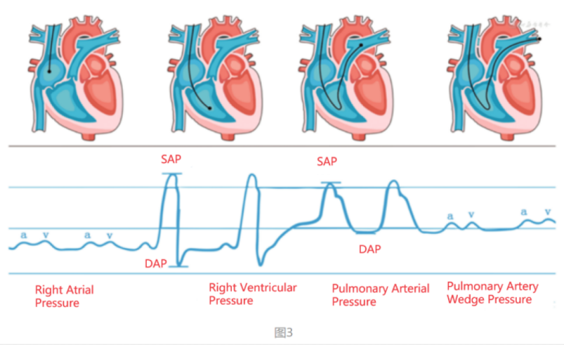
Pulmonary artery wedge pressure (PAWP) Since the valve is not present between the pulmonary vein and the left atrium, the left atrial pressure can be inversely transmitted to the pulmonary capillaries through the pulmonary vein.PAWP can estimate the pulmonary circulation status and left ventricular function, identify cardiac or pulmonary pulmonary edema, determine the therapeutic effect of vasoactive drugs, diagnose hypovolemia, and determine the effect of blood transfusion, infusion, etc. During the placement of the pulmonary artery catheter (PAC), continuous pressure changes can be recorded with the position of the catheter end (Figure 3).
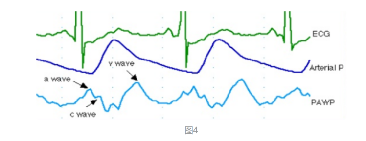
The pulmonary artery catheter can be placed about 20 cm through the internal jugular vein, the tube end reaches the right atrium, and recorded in the low flat section of the right chamber pressure waveform; the balloon is inflated, the PAC flows into the right ventricle through the tricuspid valve.When the catheter tip reaches the right ventricle, the pressure rises sharply, then the descending branch quickly drops down to near zero, when the typical right ventricular waveform (RVP) will appear. When the implantation depth reached 35 cm, the catheter entered the pulmonary artery (PAP).At this time, the systolic blood pressure changed little, but the diastolic blood pressure increased significantly, which is larger than the right ventricular diastolic pressure, manifested as the pulmonary artery pressure waveform.By advancing the catheter, the pulmonary artery branch is embedded and the PAWP waveform appears (Figure 4).
Interpretation of the PAWP waveform:
Comparison of CVP and PAWP
If you want to learn more details about our products, please check this link below: https://www.chinautech.com/products/um2017-patient-monitor.html
|


Determinants of central venous pressure
High Blood Pressure in Children
Related Article